
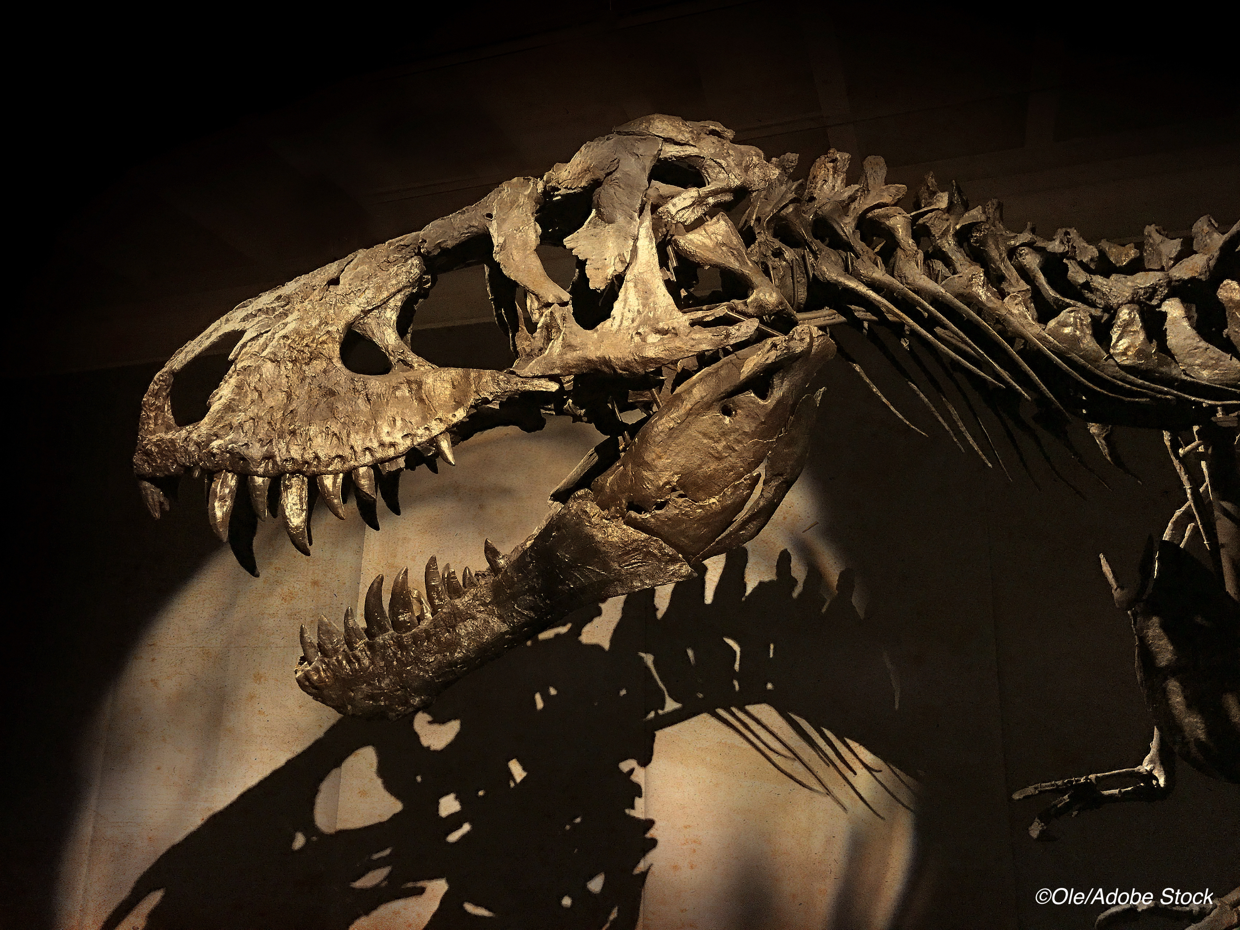
CHICAGO—When ’Tristan Otto’—one of the world’s most complete Tyrannosaurus rex skeletons—died 68 million years ago, it is likely he wasn’t a very happy camper, because researchers using 21st Century technology believe he had a truly massive toothache.
Employing dual-energy computed tomography (DECT) and with permission from the Museum für Naturkunde Berlin, Charlie Hamm, of Charite University Hospital, Berlin, and colleagues were able to determine that a curious lesion on the jawbone of Tristan Otto was the result of a bone infection.
“We hypothesized that DECT could potentially allow for quantitative noninvasive element-based material decomposition and thereby help paleontologists in characterizing unique fossils,” Hamm said at the annual meeting of the Radiological Society of North America.
The computer-assisted tomography (CT) technique enabled the researchers to overcome the difficulties of scanning a large portion of Tristan Otto’s lower jaw called the left dentary. The jawbone’s high density was particularly challenging, as CT imaging quality is known to suffer from artifacts, or misrepresentations of tissue structures, when looking at very dense objects.
“The left dentary showed two notable findings on visual inspection and CT imaging,” Hamm reported, “a diffuse thickening of nearly the entire left dentary with a homogenous distribution of calcium and a focal exophytic mass on the ventral surface of the dentary with a significant accumulation of fluorine (P<0.0001). Furthermore, the focal exophytic mass showed diminutive diffuse lucencies extending from the surface to the tooth root of the 5th replacement tooth and demonstrated a tapering shape with a fistular-like center, which also demonstrated a significant fluorine accumulation respectively (P<0.0001). The perseverance of anatomical structures within the mass suggests the diagnosis of tumefactive osteomyelitis.”
Tristan Otto was discovered by a commercial paleontologist working in Carter County, Montana. The fossilized skeleton dates back to the Late Cretaceous period. It was sold to an investment banker, who dubbed it “Tristan Otto” before loaning it out to the Museum für Naturkunde Berlin in Germany. It is one of only two original T. rex skeletons in Europe.
Hamm noted that previously, fossil studies have mostly relied on invasive sampling and analysis. DECT employs a clinical CT scanner which deploys X-rays at two different energy levels to provide information about tissue composition and disease processes not possible with single-energy CT. “We needed to adjust the CT scanner’s tube current and voltage in order to minimize artifacts and improve image quality,” Hamm said.
“While this is a proof-of-concept study, noninvasive DECT imaging that provides structural and molecular information on unique fossil objects has the potential to address an unmet need in paleontology, avoiding defragmentation or destruction,” Hamm said.
“The DECT approach has promise in other paleontological applications, such as age determination and differentiation of actual bone from replicas,” added Oliver Hampe, PhD, a paleontologist from the Museum für Naturkunde Berlin. “The experimental design, including the use of a clinical CT scanner, will allow for broad applications.”
Hamm and colleagues also collaborated with paleontologists from the Chicago’s Field Museum and colleagues from the Richard and Loan Hill Department of Biomedical Engineering at the University of Illinois at Chicago to perform a CT analysis of the world-famous T. rex “Sue” that is housed in the museum.
“With every project, our collaborative network grew and evolved into a truly multidisciplinary group of experts in geology, mineralogy, paleontology, and radiology, emphasizing the potential and relevance of the results to different scientific fields,” Hamm said.
In commenting on the study, Elliott Fishman, MD, professor of radiology, of oncology, and of surgery at Johns Hopkins University, Baltimore, told BreakingMED, “It is always interesting, when you have technology such as DECT, to use it on animals, which is commonly done in veterinary clinics, but it also gives you the opportunity to look at things like Inca mummies—which we did years ago and found out that one mummy had suffered a skull fracture—and fossils.
“With imaging, there are a lot of secrets you can learn outside of patient care,” he added. “There is always extra time to find these things, and one of the good things about imaging is that it is non-destructive testing, and we can do it very elegantly.”
John McKenna, Associate Editor, BreakingMED™
The authors had no relevant disclosures.
Fishman disclosed relationships with Seimen’s and GE.
Cat ID: 235
Topic ID: 98,235,438,791,190,481,96,235