
Photo Credit: VectorMine
The following is a summary of “Evaluation of pressure-induced pain in patients with disorders of consciousness based on functional near infrared spectroscopy,” published in the April 2025 issue of Frontiers in Neurology by Zhang et al.
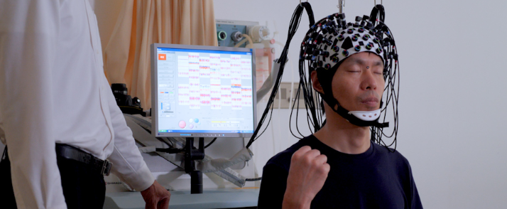
Researchers conducted a retrospective study to explore the brain’s hemodynamic responses (HRO) and functional connectivity in patients with disorders of consciousness (DoC) during acute pressure pain stimulation using near-infrared spectroscopy (NIRS).
They assessed patients diagnosed with DoC using pressure stimulation while measuring brain activity with NIRS. Changes in oxygenated hemoglobin (HbO) and deoxygenated hemoglobin (HbR) concentrations were monitored across regions of interest (ROIs): primary somatosensory cortex (PSC), primary motor cortex (PMC), dorsolateral prefrontal cortex (dPFC), somatosensory association cortex (SAC), temporal gyrus (TG), and frontopolar area (FPA). Functional connectivity was evaluated during pre-stimulation, stimulation, and post-stimulation phases.
The results showed no significant changes in HbO or HbR concentrations during stimulation vs. baseline or stimulation vs. post-stimulation, indicating minimal activation of targeted brain regions. However, functional connectivity between the PSC, PMC, and dPFC significantly enhanced during stimulation (r > 0.9, P < 0.001), suggesting greater coordination among sensory, motor, and cognitive regions.
Investigators found minimal pain-induced brain activation in patients with DoC, but enhanced functional connectivity during pain stimulation suggested the brain continued processing pain through coordinated regional activity. The findings highlighted the importance of assessing functional connectivity to evaluate pain processing in these patients.
Source: frontiersin.org/journals/neurology/articles/10.3389/fneur.2025.1542691/full


Create Post
Twitter/X Preview
Logout